
Coronary Circulation Animation | Systole vs Diastole, Regulation & Oxygen Demand: Dr G Bhanu Prakash
📌 𝐅𝐨𝐥𝐥𝐨𝐰 𝐨𝐧 𝐈𝐧𝐬𝐭𝐚𝐠𝐫𝐚𝐦:- https://www.instagram.com/drgbhanuprakash 📢 𝗝𝗢𝗜𝗡 𝗢𝗨𝗥 𝗢𝗙𝗙𝗜𝗖𝗜𝗔𝗟 𝗧𝗘𝗟𝗘𝗚𝗥𝗔𝗠 𝗖𝗛𝗔𝗡𝗡𝗘𝗟𝗦 👨⚕️ 𝗗𝗿. 𝗚. 𝗕𝗵𝗮𝗻𝘂 𝗣𝗿𝗮𝗸𝗮𝘀𝗵 – 𝗢𝗳𝗳𝗶𝗰𝗶𝗮𝗹 𝗧𝗲𝗹𝗲𝗴𝗿𝗮𝗺 𝗖𝗵𝗮𝗻𝗻𝗲𝗹 🔗 https://t.me/bhanuprakashdr 🎯 NEET PG 2027 Core Revision– 𝗘𝘅𝗰𝗹𝘂𝘀𝗶𝘃𝗲 𝗧𝗲𝗹𝗲𝗴𝗿𝗮𝗺 𝗖𝗵𝗮𝗻𝗻𝗲𝗹 🔗 https://t.me/NEET_PG_MED 🚀 𝗙𝗠𝗚𝗘 – 𝗘𝘅𝗰𝗹𝘂𝘀𝗶𝘃𝗲 𝗧𝗲𝗹𝗲𝗴𝗿𝗮𝗺 𝗖𝗵𝗮𝗻𝗻𝗲𝗹 🔗 https://t.me/FmgExams 📚 𝗠𝗕𝗕𝗦 𝗨𝗻𝗶𝘃𝗲𝗿𝘀𝗶𝘁𝘆 𝗘𝘅𝗮𝗺𝘀 – 𝗘𝘅𝗰𝗹𝘂𝘀𝗶𝘃𝗲 𝗧𝗲𝗹𝗲𝗴𝗿𝗮𝗺 𝗖𝗵𝗮𝗻𝗻𝗲𝗹 🔗 https://t.me/MbbsExams 🩺 𝗣𝗮𝗿𝗮𝗺𝗲𝗱𝗶𝗰𝗮𝗹 𝗘𝘅𝗮𝗺𝘀 – 𝗘𝘅𝗰𝗹𝘂𝘀𝗶𝘃𝗲 𝗧𝗲𝗹𝗲𝗴𝗿𝗮𝗺 𝗖𝗵𝗮𝗻𝗻𝗲𝗹 🔗 https://t.me/paramedicalexams 🫀 **Coronary Blood Flow Animation – Regulation, Myocardial Perfusion & Coronary Circulation | Dr G Bhanu Prakash** 🫀 **Coronary Blood Flow – Core Concept** The myocardium receives its blood supply through the coronary circulation. The right and left coronary arteries arise from the aortic root and deliver oxygenated blood to cardiac muscle. Because the myocardium has a very high metabolic requirement and already extracts a large proportion of oxygen from coronary arterial blood at rest, increases in myocardial oxygen demand are met predominantly by **increasing coronary blood flow rather than substantially increasing oxygen extraction**. 📈 **Why Is Coronary Blood Flow Greater During Diastole?** Coronary perfusion changes throughout the cardiac cycle. This is particularly important in the **left ventricular myocardium**. During systole, contracting ventricular muscle compresses intramyocardial coronary vessels, especially in the subendocardial region, thereby increasing vascular resistance and reducing left coronary blood flow. During diastole, myocardial relaxation reduces this extravascular compression and allows coronary perfusion to increase. Therefore, **left coronary blood flow is predominantly diastolic**. ❤️ **Right vs Left Coronary Blood Flow** The right ventricle generates considerably lower intramyocardial pressure than the left ventricle. Consequently, compression of right coronary vessels during systole is less pronounced, allowing the right coronary circulation to maintain relatively greater flow during systole compared with the left coronary circulation. Understanding this difference is important for interpreting coronary physiology and myocardial ischemia. 🧠 **Regulation of Coronary Blood Flow** Coronary circulation is regulated mainly according to the metabolic requirements of the myocardium. Increased cardiac activity raises myocardial oxygen consumption and promotes local vasodilation, thereby increasing coronary blood flow. Important local metabolic influences include reduced tissue oxygen availability and accumulation or signaling of vasodilator metabolites such as **adenosine**, along with contributions from carbon dioxide, hydrogen ions, potassium, nitric oxide, and other local regulatory mechanisms. ⚡ **Myocardial Oxygen Demand** Major determinants of myocardial oxygen consumption include **heart rate, myocardial contractility, and ventricular wall stress**. An increase in any of these factors can increase cardiac oxygen demand. Coronary vasodilation normally increases blood flow to match this increased metabolic requirement. When coronary blood supply cannot adequately meet myocardial oxygen demand, **myocardial ischemia** can develop. 🩸 **Coronary Perfusion Pressure** Coronary blood flow depends on the pressure gradient across the coronary vascular bed and coronary vascular resistance. For the left ventricular myocardium, coronary perfusion is strongly influenced by **aortic diastolic pressure and left ventricular diastolic pressure**. A reduction in aortic diastolic pressure or an increase in ventricular filling pressure can compromise effective coronary perfusion, particularly in vulnerable subendocardial regions. ⏱️ **Why Tachycardia Can Reduce Coronary Perfusion** As heart rate increases, diastole becomes disproportionately shorter. Because left ventricular coronary perfusion occurs predominantly during diastole, marked tachycardia reduces the available coronary filling time while simultaneously increasing myocardial oxygen consumption. This combination explains why tachycardia can aggravate ischemia in patients with significant coronary artery disease. 🚨 **Why Is the Subendocardium Vulnerable to Ischemia?** The subendocardial myocardium experiences high compressive forces during ventricular contraction and is therefore particularly dependent on adequate diastolic perfusion. Conditions that reduce coronary perfusion pressure, shorten diastole, increase ventricular wall stress, or restrict coronary blood supply can preferentially compromise the subendocardial region. #CoronaryBloodFlow #CoronaryCirculation #CoronaryPerfusion #CardiacPhysiology #CardiovascularPhysiology #HeartPhysiology #CoronaryArtery #MyocardialPerfusion #MyocardialOxygenDemand #CoronaryArteryDisease #MyocardialIschemia #MedicalAnimation #MedicalEducation #Physiology #Cardiology #MedicalStudents #USMLE #USMLEStep1 #USMLEStep2CK #NBME #ShelfExam #COMLEX #COMLEXUSA #MCCQE #UKMLA #PLAB #MRCP
Suggested videos
Sciatic Nerve Anatomy Animation - Course, Relations, Branches, Distribution & Clinical anatomy
Dr.G Bhanu Prakash Animated Medical Videos · 6:56
Limbic System Anatomy Animation🧠 Components, Functions & Clinical Relevance | Dr G Bhanu Prakash
Dr.G Bhanu Prakash Animated Medical Videos and Conceptual Minds · 7:43

Anatomy of Abdomen Rapid Revision | Dr krishna Sahith
Dr.G Bhanu Prakash Animated Medical Videos · 6:47:37
Anatomy of Head Rapid Revision - MBBS & FMGE
Dr.G Bhanu Prakash Animated Medical Videos · 3:56:02

Neuroanatomy Rapid Revision : Dr Krishna Sahith
Dr.G Bhanu Prakash Animated Medical Videos · 7:50:16
Anatomy of Stomach Animation: Blood supply, Venous drainage, Nerve supply, Histology & Embryology
Dr.G Bhanu Prakash Animated Medical Videos · 23:32

Remarkable Rapid Revision Internal Medicine (FMGE 2026) | Dr. Sandeep Sharma
Dr.G Bhanu Prakash Animated Medical Videos · 9:42:44
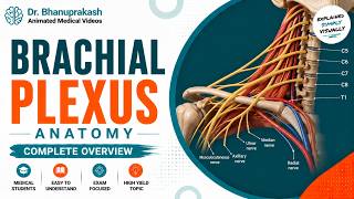
The Brachial Plexus Anatomy USMLE: Roots, Trunks, Divisions, Cords, Branches, Clinical anatomy
Dr.G Bhanu Prakash Animated Medical Videos · 15:12